The electricity of food
The metabolic pathways of the macronutrients and how mitochondria reduce food to a source of electrons
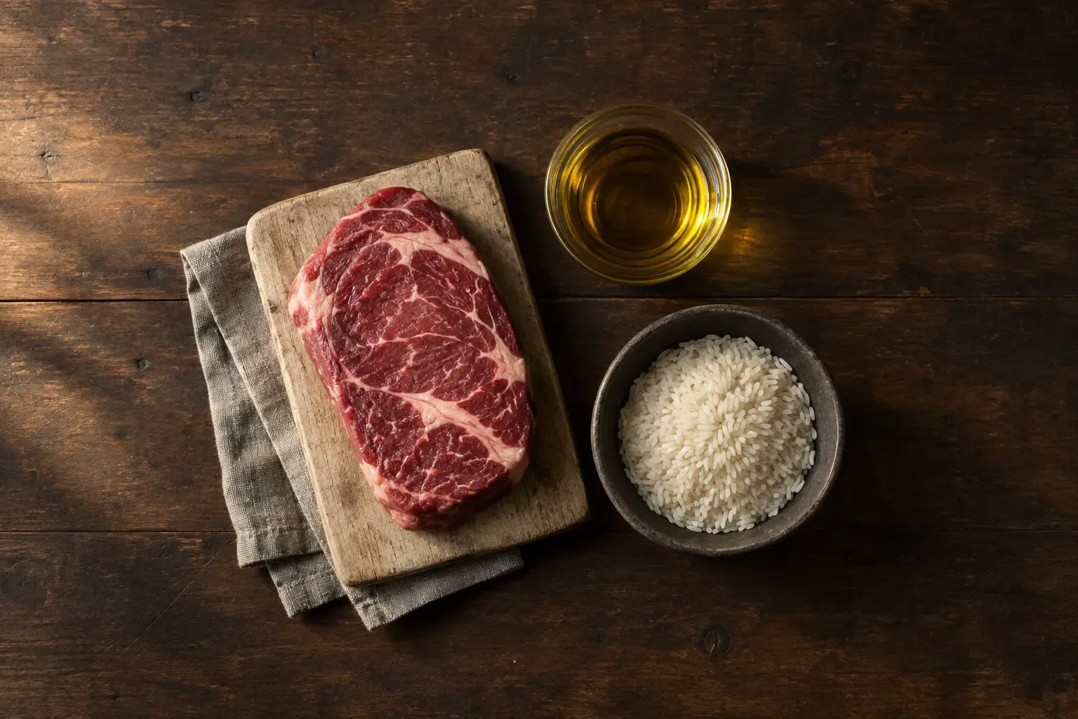
Behind every plate of food there is a biological reality not everyone knows about. A cut of meat, a splash of olive oil or a bowl of rice are, above all, collections of organic molecules held together by chemical bonds. The main job of metabolism is not to use those molecules as they arrive through the mouth, but to take them apart step by step in order to extract something far more elemental: their electrons.
Inside almost every one of your cells there are hundreds of power plants called mitochondria. Their mission is to collect those high-energy electrons using organic molecules that act as “taxis” (NADH and FADH₂). These taxis carry the electrons to a microscopic motorway set into the inner mitochondrial membrane: the electron transport chain.
As the electrons hop from one station to the next inside the mitochondrion, they release energy. The cell uses that movement to pump protons out of the mitochondrial matrix, creating a microscopic “hydroelectric dam”. When those protons flow back in through a molecular turbine called ATP synthase, the body’s energy currency is generated: ATP (adenosine triphosphate).
For this electrical circuit not to collapse, a final acceptor is needed to collect the electrons at the end of the run. That role is played by the oxygen you breathe, which combines with the leftover electrons and protons to form something as simple as water (H₂O). Without oxygen the current stops, ATP production grinds to a halt and the cell is left with no energy.
The three metabolic pathways
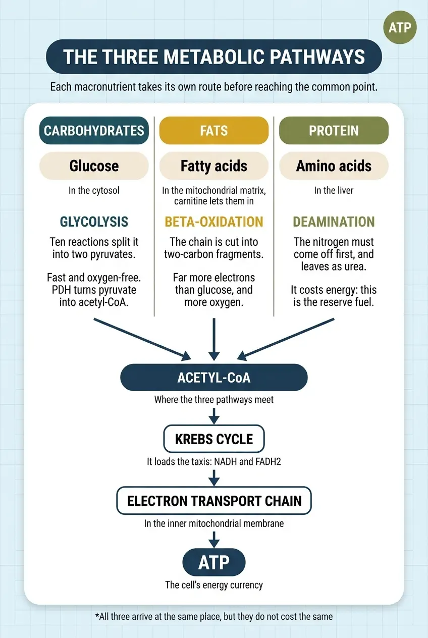
Although the final goal is always to extract electrons in order to make ATP, the way the body processes each macronutrient follows radically different biochemical routes before reaching a common point: the acetyl-CoA molecule.
Carbohydrates: the glycolysis pathway
Carbohydrates are broken down into glucose. Inside the cellular fluid (the cytosol), glucose undergoes glycolysis, a series of ten chemical reactions that split it into two molecules of pyruvate.
- Speed without oxygen. Its great advantage is that it can generate ATP very quickly and with no need for oxygen. It is the immediate resource for intense efforts.
- The step into the mitochondrion. To extract the rest of its energy, pyruvate enters the mitochondrion, where the enzyme pyruvate dehydrogenase (PDH) converts it into acetyl-CoA to feed the Krebs cycle and the electron chain.
Fats: the beta-oxidation pathway
Fats are broken down into fatty acids. To be used, they must travel into the mitochondrion with the help of a special transport called carnitine.
Once inside they undergo beta-oxidation: a process in which the fat chain is cut repeatedly into two-carbon fragments, generating an enormous amount of acetyl-CoA and of taxis loaded with electrons (NADH and FADH₂). Because fats are packed with carbon-hydrogen bonds, they contain far more electrons than carbohydrates do. They need more oxygen to “burn”, but they offer an almost inexhaustible energy reserve.
Protein: the deamination pathway
Proteins are made of amino acids. Unlike fats and carbohydrates, amino acids contain nitrogen in an amino group (-NH₂). Since nitrogen is of no use for generating energy and is toxic in excess, the liver has to remove it first through deamination.
The nitrogen removed is turned into urea to be excreted in the urine. Only then can the remaining carbon skeleton be converted into intermediates for making ATP. For that reason, using protein as fuel carries an extra metabolic cost, and the body only resorts to it secondarily or in situations of extreme need.
Metabolic flexibility and rigidity
The body does not pick its fuel at random. The choice depends on hormonal state and on energy demand.
- The postprandial state (after eating). The rise in blood glucose prompts the beta cells of the pancreas to secrete insulin. Insulin activates phosphatases that stimulate glycogen synthase and the enzyme phosphofructokinase-1 (PFK-1), promoting the storage and oxidation of glucose. At the same time it inhibits hormone-sensitive lipase (HSL) in adipose tissue, blocking the release of fatty acids into the circulation. In other words, stored fat is kept from leaving: while insulin is high, the body prioritises using and storing glucose.
- The fasting or exercising state. A drop in blood glucose or a rise in physiological stress stimulates the secretion of glucagon and catecholamines (adrenaline and noradrenaline). These hormones activate HSL through the cyclic AMP (cAMP) pathway, mobilising fatty acids for oxidation.
- Exercise intensity. During physical activity, muscle contraction activates the enzyme AMPK (AMP-activated protein kinase) because of the ATP being consumed. At low or moderate intensities, such as walking, the presence of oxygen allows beta-oxidation to cover most of the demand. As intensity rises, the recruitment of fast-twitch muscle fibres and the limited rate at which fatty acids can be transported into the mitochondrion shift substrate selection towards glycolysis, regardless of insulin concentration.
What is metabolic rigidity?
If metabolic flexibility is a healthy body’s ability to switch smoothly between burning fat and burning glucose, rigidity appears when that capacity to adapt breaks down in either direction:
- An inability to burn fat. Sustaining constant insulin peaks through caloric excess and a sedentary life keeps access to fat blocked. The cell turns “rigid” and depends continuously on an external supply of sugar in order to function.
- A temporary inability to process glucose. Conversely, if someone removes carbohydrates entirely for a long time, the enzyme pyruvate dehydrogenase (PDH) lowers its activity. If that person suddenly eats a large amount of carbohydrate, their body will take a while to restart the glycolytic machinery and will show transiently high glucose peaks.
A practical strategy: movement and the quality of your fats
Two factors are decisive in daily life if this metabolic machinery is to keep working properly: muscular stimulus and the quality of the fuel.
The problem with sedentary living
A sedentary life reduces mitochondrial density and insulin sensitivity. Exercise, by contrast, acts as a direct switch. When muscles contract, the enzyme AMPK detects the energy being spent and prompts the GLUT4 transporters to travel to the cell surface to capture glucose from the blood with no need for insulin.
The stimulus required to maintain this machinery does not demand elite training: strength or moderate endurance sessions spread across the week, or simply accumulated daily activity, are enough to promote mitochondrial biogenesis. You do not have to be a professional athlete to keep sound metabolic flexibility, but physical exercise is fundamental to a healthy body.
The chemical stability of fats
Fats are an important source of energy, but not all lipids behave the same way in the body. The key lies in their molecular structure:
- Saturated and monounsaturated fats. Saturated fats (found in the meat of pasture-raised animals, in lard or in coconut oil) and monounsaturated ones (such as the oleic acid in olive oil) have either no double bonds or only one. That structure makes them highly stable and resistant to oxidation. Quality animal fats also supply fat-soluble vitamins (A, D₃ and K₂) and coenzyme Q₁₀, which is key to mitochondrial performance.
- Polyunsaturated fats (PUFAs) and antinutrients. Refined vegetable oils and some nuts in excess contain high amounts of polyunsaturated fats (omega-6), whose multiple double bonds leave them vulnerable to being altered by heat or oxidation. Raw nuts also contain phytic acid and lectins, compounds that can reduce the absorption of essential minerals such as zinc or magnesium.
The environmental factor in mitochondrial function: mitochondrial performance is also modulated by environmental stimuli such as sunlight. Radiation in the near-infrared range penetrates tissue and stimulates the enzyme cytochrome c oxidase — one of the stations of the electron chain — optimising ATP production. Eating indoors is not the same as eating outdoors, just as the body cannot be efficient if it is systematically deprived of sunlight.
How food policy altered our biology
If our body is such a precise machine, why have metabolic rigidity and type 2 diabetes become a modern epidemic? The answer combines scientific bias with commercial pressure.
In the 1950s, the researcher Ancel Keys pushed the hypothesis that saturated fats were solely responsible for heart disease. Although his study left out the data from countries that did not fit his theory, the idea took deep root in the institutions.
Years later, historical documents revealed that in 1965 the Sugar Research Foundation — the sugar industry lobby in the United States — secretly paid scientists at Harvard University to publish reviews that played down the impact of sugar on cardiovascular health and pointed to fats as the only enemy. The definitive outcome of that process came in 1992, when the United States Department of Agriculture (USDA) launched the world-famous food pyramid.
Heavily influenced by the grain industry, the guide put at its base the recommendation to eat numerous daily servings of bread, pasta, rice and flour. Brands responded by flooding the shelves with low-fat products and, to make up for the loss of flavour once the fat was gone, added large amounts of refined sugars and fructose syrups. Three decades after that nutritional experiment, rates of obesity and metabolic disease reached historic highs across the world.
No macronutrient is harmful in itself
The human body does not understand fashions or dietary dogma: it responds to the principles of biochemistry, organic chemistry and thermodynamics, and it is perfectly equipped to metabolise all three macronutrients.
Understanding that food is a source of electrons destined to produce ATP lets us see nutrition far more clearly. It is not about demonising macronutrients, but about giving the mitochondria chemically stable food, keeping muscle mass active through movement, and giving the body back the capacity to adapt it was designed with.
References and scientific support
- The reference textbook on bioenergetics. Nicholls, D. G., & Ferguson, S. J. (2013). Bioenergetics 4. Academic Press. View the book on ScienceDirect
- The chemiosmotic theory that explained how ATP is made. Mitchell, P. (1961). Coupling of phosphorylation to electron and hydrogen transfer by a chemi-osmotic type of mechanism. Nature, 191(4784), 144-148. View study (DOI)
- Fuel selection when the switch fails. Kelley, D. E., & Mandarino, L. J. (2000). Fuel selection in human skeletal muscle in insulin resistance: a reexamination. Diabetes, 49(5), 677-683. View study (DOI)
- What metabolic flexibility actually is. Goodpaster, B. H., & Sparks, L. M. (2017). Metabolic flexibility in health and disease. Cell Metabolism, 25(5), 1027-1036. View study on PubMed Central
- The Randle cycle: glucose and fat competing for the same furnace. Randle, P. J., Garland, P. B., Hales, C. N., & Newsholme, E. A. (1963). The glucose fatty-acid cycle. Its role in insulin sensitivity and the metabolic disturbances of diabetes mellitus. The Lancet, 281(7285), 785-789. View study (DOI)
- Muscle contraction takes up glucose without insulin. Richter, E. A., & Hargreaves, M. (2013). Exercise, GLUT4, and skeletal muscle glucose uptake. Physiological Reviews, 93(3), 993-1017. View study (DOI)
- AMPK as the cell’s energy sensor. Hardie, D. G., Ross, F. A., & Hawley, S. A. (2012). AMPK: a nutrient and energy sensor that maintains energy homeostasis. Nature Reviews Molecular Cell Biology, 13(4), 251-262. View study on PubMed Central
- Red and infrared light stimulate cytochrome c oxidase. Karu, T. I. (2008). Mitochondrial signaling in mammalian cells activated by red and near-IR radiation. Photochemistry and Photobiology, 84(5), 1091-1099. View study (DOI)
- What happens to a polyunsaturated oil when it is heated. Grootveld, M., Silwood, C. J. L., Addis, P., Claxson, A., Serra, B. B., & Viana, M. (2001). Health effects of oxidized heated oils. Foodservice Research International, 13(1), 41-55. View study (DOI)
- Phytic acid and mineral absorption. Schlemmer, U., Frølich, W., Prieto, R. M., & Grases, F. (2009). Phytate in foods and significance for humans: food sources, intake, processing, bioavailability, protective role and analysis. Molecular Nutrition & Food Research, 53(S2), S330-S375. View study (DOI)
- The sugar industry’s internal documents. Kearns, C. E., Schmidt, L. A., & Glantz, S. A. (2016). Sugar industry and coronary heart disease research: a historical analysis of internal industry documents. JAMA Internal Medicine, 176(11), 1680-1685. View study on PubMed Central
- The study that put saturated fat in the dock. Keys, A. (1980). Seven Countries: A Multivariate Analysis of Death and Coronary Heart Disease. Harvard University Press. View the study’s publications
- How industry shapes dietary guidelines. Nestle, M. (2013). Food Politics: How the Food Industry Influences Nutrition and Health. University of California Press. View the book at the publisher
We're building the space for you to comment on the articles.
Continue reading

The molecular architecture
Chain length, degree of saturation and the metabolic fate of fatty acids

Glycine: the forgotten building block of life
The key amino acid that slows cellular ageing, improves sleep and rebuilds your tissues from within